We use cookies and similar tracking technologies to enhance your browsing experience, analyze website traffic, and provide personalized content and advertisements. Some cookies are essential for the proper functioning of our website, while others help us improve performance, enable additional features, or deliver tailored marketing. By clicking “Accept All Cookies,” you consent to the use of all cookies and the sharing of this information with our partners. You can manage your cookie preferences at any time by clicking “Cookies Settings” below.
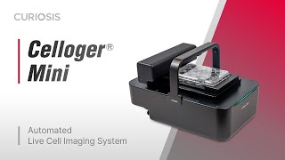
Celloger® Mini
- Discontinued product -
Celloger Mini is an automated live cell imaging system based on bright-field microscopy with fully motorized stages. This all-around and compact system provides autofocusing, time lapse imaging, and analytical software that lets you perform various types of sophisticated researches.
Diversify your research paradigm with our integrated live cell imaging system, Celloger Mini
Remotely monitor live cells inside the incubator without disturbing the environment suitable for cell culture. You can monitor cells in real-time or with the time-lapse function, cell images are captured automatically for days or even weeks without having to move the cells in and out of the incubator.
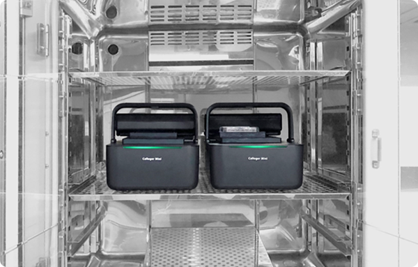
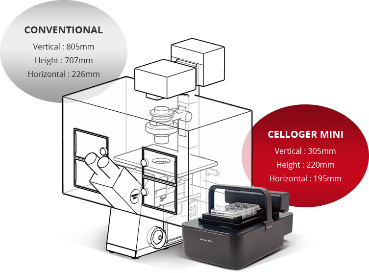
Compact size
Compact size makes it straightforward to install and handle, so there is no need for calibration and complicated maintenance procedures. Small-in-size also provides a room for space utilization inside incubators making it possible for multiple unit installation.
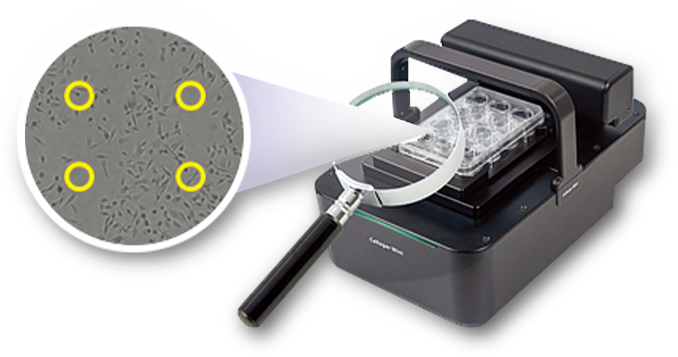
Multi-point imaging
Capturing up to 999 positions
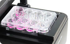
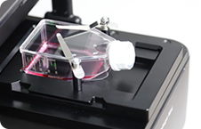
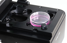
The moving XY stage allows imaging of multiple positions within the travel range of 117 x 77mm and it is even possible to capture multiple points within a well. Also, cells cultured in different vessel types such as well plates, flasks, and dishes can be imaged.
Well-plate
Culture flask
Culture dish
User-friendly functions
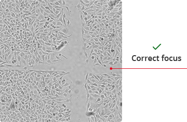
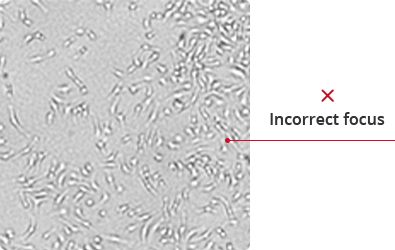
Autofocus
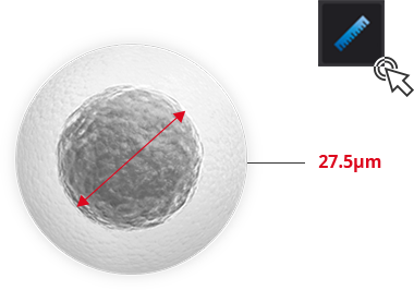
Ruler
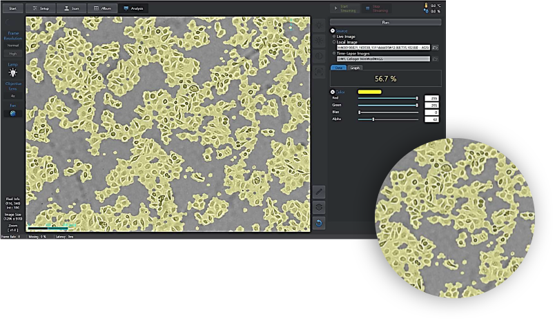
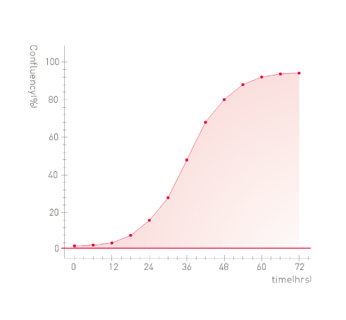
Cell confluency & growth curve
User-friendly functions
Cell
Cell type
Species
Tissue
MDA-MB-231
Epithelial cell
Human
Breast
HeLa
Epithelial cell
Human
Cervix
AGS
Epithelial cell
Human
Stomach
HCT116
Epithelial cell
Human
Colon
HaCaT
Epithelial cell
Human
Skin(Keratinocyte)
MCF-7
Epithelial cell
Human
Breast
SH-SY5Y
Epithelial cell
Human
Bone marrow
LoVo
Epithelial cell
Human
Colon
A549
Epithelial cell
Human
Lung
293T
Epithelial cell
Human
Kidney
CPAE
Endothelial cell
cattle
Pulmonary artery
NIH3T3
Fibroblast
mouse
Embryo
Hs27
Fibroblast
Human
Skin
Raw264.7
Macrophage
mouse
Blood(ascites)
Specification
Dimension
195 x 305 x 220 mm
Weight
4.5kg / 9.9lb
Objective Lens
4X
Imaging modes
Bright field
Camera
1.25MP / 5MP CMOS
Resolution
1296 x 970 / 2592 x 1942 pixel
Stage
Motorized XYZ
Imaging positions
Multiple
File export format
TIFF, JPEG, PNG, AVI
Culture vessels
Flask, dish, well plate, slide
Operating environment
5~40℃, 20~95% humidity
Power requirements
100-240V, ~50/60Hz
Ordering information
Catalog No.
Description
CRCLG-MB01
Celloger Mini, Live cell imaging system (Bright Field)


.png)


 Celloger® Mini